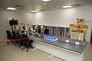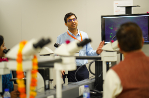
In a groundbreaking advancement for diagnostic capabilities and medical education, Department of Pathology, in collaboration with the Department of Microbiology, unveiled the Deca-Head Microscope. This state-of-the-art technology is set to significantly enhance diagnostic precision and foster interdisciplinary collaboration in both clinical and educational settings.
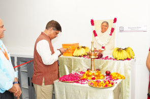
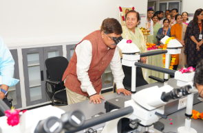
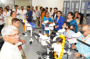
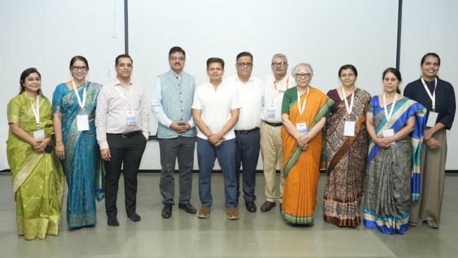
The event was led by Dr. Sanjeev Singh, Medical Director of Amrita Hospital, Faridabad, along with Mrs. Varsha, Dr. K. R. Rathi, and Dr. Anubhav Pandey. The unveiling was attended by esteemed faculty members from Pathology, Microbiology, Oncology, and other departments, as well as hospital staff, who gathered to celebrate this remarkable milestone.
The Deca-Head Microscope is designed to improve diagnostic accuracy by allowing real-time discussions and collaborative assessments across disciplines. The technology is equipped with high-resolution optics that enhance the clarity of histopathological specimen evaluation. Its ergonomic and modular design ensures user comfort during prolonged use, while the microscope’s broadcasting facility enables the live sharing of case studies on online platforms, facilitating remote collaboration.
This innovative tool is expected to make a significant impact on patient care by aiding in the diagnosis of complex cases, including rare CNS tumors and challenging biopsies. With its capability for real-time intra-departmental consultations, the microscope will help reduce turnaround times, enabling quicker clinical decision-making. Furthermore, the technology promotes better clinicopathological integration, aligning histological interpretations with clinical profiles for more effective treatment planning.
In addition to improving patient care, the microscope will serve as an educational hub for residents and postgraduate trainees. It allows for interactive learning sessions and live demonstrations, enhancing engagement and comprehension. The Deca-Head Microscope will also play a crucial role in enriching clinicopathological meetings by providing a central platform for multidisciplinary dialogue between clinicians and pathologists. It streamlines the process of capturing images for research and documentation purposes, further contributing to the hospital’s ongoing academic efforts. The microscope’s role in collaborative patient management, through morphology-informed decision-making, underscores its importance in advancing holistic patient care.