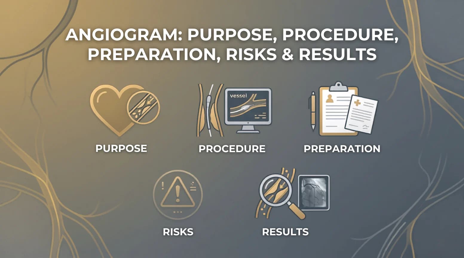
An angiogram is a diagnostic imaging test that allows doctors to examine the blood vessels in your heart, brain, kidneys, legs, or other parts of the body. Using a contrast dye and X-ray imaging, the procedure highlights areas of narrowing, blockage, or abnormal blood flow that cannot be detected through standard scans. It remains one of the most reliable methods for identifying vascular conditions and guiding the next steps in treatment.
Doctors recommend an angiogram when a patient shows symptoms of poor blood flow, such as chest pain, breathlessness, or numbness in the limbs, or when other tests point toward a possible blockage. This guide covers what an angiogram is used for, how the procedure works, how to prepare for it, and what to expect in terms of risks and results.
What Is an Angiogram?
An angiogram, also called angiography, is a medical imaging test that shows how blood moves through the arteries and veins in your body. During the procedure, a doctor injects a contrast dye into the bloodstream through a thin, flexible tube called a catheter, making the blood vessels visible on X-ray, CT, or MRI imaging. This allows doctors to locate problems that cannot be picked up through standard imaging or physical exams.
- Reveals narrowed or blocked arteries caused by plaque buildup
- Detects aneurysms and weak spots in blood vessel walls
- Identifies abnormalities in blood vessels, such as aneurysms or abnormal blood flow
- Guides decisions on medication, angioplasty, or bypass surgery
Why Is an Angiogram Performed?
Doctors recommend an angiogram when symptoms or earlier test results point toward a possible problem in the heart or blood vessels. It helps confirm a diagnosis, assess how severe the issue is, and guide the right course of treatment.
- You experience angina for the first time.
- Your angina is becoming more severe, occurs more frequently, does not improve, or happens even when you are resting, which may indicate unstable angina.
- You have aortic stenosis or another heart valve disorder.
- You have unusual or atypical chest pain despite normal results from other tests.
- You have received an abnormal result on a cardiac stress test.
- You are scheduled for heart surgery and have a high risk of coronary artery disease.
- You have heart failure.
- You have been diagnosed with a heart attack.
Who Needs an Angiogram?
Not everyone with heart or blood vessel concerns needs an angiogram. Doctors weigh a few factors before recommending it.
Symptoms that prompt evaluation:
- Chest pain, breathlessness, irregular heartbeat, or leg pain during activity
Risk factors doctors consider:
- High blood pressure, high cholesterol, diabetes, smoking, or a family history of heart disease
When it may not be recommended:
- Low risk of heart attack or angina well controlled with medication
- Severe kidney disease, a previous serious reaction to contrast media, or certain bleeding disorders, where alternative imaging or additional precautions may be considered
Types of Angiograms
The type of angiogram a doctor recommends depends on which part of the body needs to be examined, how detailed the images need to be, and whether the situation calls for a diagnostic look, an immediate treatment option, or both.
Coronary Angiogram
This is the most common type of angiogram and is used to examine the arteries that supply blood to the heart. A catheter is guided through a blood vessel in the wrist or groin up to the coronary arteries, where contrast dye is injected. X-ray imaging then captures how the dye moves, revealing blockages, narrowing, or plaque buildup. If a blockage is found, doctors can often treat it in the same session through angioplasty or stenting.
CT Coronary Angiogram
This is a non-invasive alternative that combines a CT scanner with contrast dye, injected through an IV rather than a catheter. The imaging portion is typically brief and provides detailed images of the coronary arteries without the need for catheter insertion into the heart arteries. It may be used to evaluate patients with stable chest pain when appropriate based on their symptoms, risk level, and clinical assessment. Unlike invasive coronary angiography, it does not allow doctors to treat a blockage during the scan.
Peripheral Angiogram
This looks at blood flow in the arms and legs to identify blockages or narrowing associated with peripheral artery disease. It follows the same catheter-based approach as a coronary angiogram, with the catheter guided to the arteries in the affected limb, and can help doctors plan treatments such as angioplasty for improving circulation.
Cerebral Angiogram
This focuses on the blood vessels in the brain and is used to detect aneurysms, blockages, arteriovenous malformations, or other abnormalities linked to stroke risk. A catheter is threaded from an artery in the leg up to the blood vessels supplying the brain, and imaging is used to map blood flow in detail. The head is kept still during the procedure to ensure clear images.
Pulmonary Angiogram
This examines the blood vessels in the lungs and is most often used to diagnose a pulmonary embolism, a blockage caused by a blood clot. CT pulmonary angiography is now more commonly used to diagnose pulmonary embolism because it is less invasive and widely available. Conventional pulmonary angiography is generally reserved for selected cases, including situations where catheter-based treatment may also be considered.
Angiogram vs Other Heart Tests
An angiogram is not always the first test a doctor orders. Depending on a patient's symptoms and risk level, doctors often start with a less invasive test and move to an angiogram only if the results point toward a blockage that needs a closer look or immediate treatment.
Diagnostic Tests: An Overview
A Coronary Angiogram is an invasive test that provides a direct, real-time view of blockages inside the coronary arteries. It is best used for confirming a diagnosis and treating a blockage in the same session.
CT Coronary Angiography is a non-invasive test that produces detailed anatomical images of the coronary arteries via a CT scan. It is typically used as a first-line test for patients with stable chest pain.
A Stress Test is also non-invasive and shows how the heart performs under physical or induced stress. It is best used for assessing whether reduced blood flow triggers symptoms during exertion.
An Echocardiogram is a non-invasive test that uses ultrasound to show heart structure, valve function, and pumping ability. It is best used for evaluating chronic conditions like heart failure or valve disease.
A Cardiac MRI, another non-invasive test, provides detailed images of heart tissue, scarring, and function. It is best used for assessing tissue damage and complex cases where other tests may fall short.
How Doctors Choose the Right Diagnostic Test?
The choice of test depends on the clinical question being asked. If a doctor needs to know whether the heart muscle is structurally sound, an echocardiogram is usually the starting point. If the concern is reduced blood flow during exertion, a stress test or cardiac MRI can help assess ischemia. When there is a strong suspicion of significant coronary artery disease, doctors may recommend invasive coronary angiography because it provides detailed images of the coronary arteries and can allow treatment, such as angioplasty or stenting, during the same procedure when appropriate.
Preparing for an Angiogram
Preparation for an angiogram usually begins a few days before the procedure, and following each step closely helps reduce the risk of complications and delays.
1. Medical Consultation
The doctor reviews the patient's full medical history, current medications, kidney function, and any past reactions to contrast dye before scheduling the procedure. This is also the time to raise questions or concerns about the test.
2. Blood Tests and Imaging
Blood tests may be performed before the procedure to assess kidney function, blood counts, or clotting status, depending on the patient's medical history, medications, and the type of angiogram planned. The procedure may be postponed if results fall outside the acceptable range, since the contrast dye is processed through the kidneys.
3. Medication Instructions
Patients taking blood thinners, diabetes medicines, or other medications may need specific instructions before the procedure. Patients should not stop or change any medication unless advised by their doctor. Diabetic patients should ask their doctor for specific instructions on insulin timing during the fasting period.
4. Fasting Requirements
Fasting requirements vary depending on the type of angiogram, sedation plan, and hospital protocol. Patients should follow the specific instructions provided by their medical team about when to stop eating and drinking before the procedure.
5. Allergy Assessment
Patients should tell their doctor if they have previously experienced a reaction to iodinated contrast media. A previous reaction does not necessarily rule out the procedure, but the medical team may take additional precautions based on the type and severity of the reaction. Shellfish allergy alone does not indicate an allergy to iodinated contrast.
What to Expect Before an Angiogram?
Will I need to be admitted to the hospital?
Most patients check in on the morning of the procedure and are treated as outpatients, going home the same day if there are no complications. Some cases may require overnight admission, depending on the hospital's protocol and the patient's health.
What happens during the consent process?
Before anything begins, the doctor explains the procedure, its risks, and any alternatives, and answers questions the patient may have. Once the patient understands and agrees, they sign a consent form confirming this.
How is the catheter insertion site prepared?
The chosen site, usually the wrist, groin, or elbow, is cleaned thoroughly and shaved if needed to lower the risk of infection. An IV line is also placed in the arm to allow the team to give medication during the procedure.
What is discussed about anaesthesia?
The team explains that local anaesthesia will be used to numb the insertion site, along with a mild sedative through the IV to help the patient stay relaxed. Patients remain awake and responsive, since they may need to follow instructions like holding their breath briefly.
What checks happen right before the procedure starts?
The medical team confirms the patient's identity, reviews the procedure plan one final time, and checks that vital signs, monitoring equipment, and the insertion site are all in order.
How Is an Angiogram Performed?
Before the Procedure
The patient changes into a hospital gown and lies down on the X-ray table. The team places an IV line in the arm, attaches ECG leads to monitor heart rhythm, and cleans and numbs the site where the catheter will be inserted, usually the wrist, groin, or elbow.
During the Procedure
A mild sedative helps the patient stay relaxed, though they remain awake throughout. The steps that follow are:
- A needle is used to access the chosen blood vessel, and a thin wire is guided through it
- A catheter is threaded over the wire and moved carefully toward the area being examined, such as the coronary arteries or brain
- Contrast dye is injected through the catheter once it reaches the target site
- X-ray imaging captures how the dye moves through the blood vessels, revealing blockages, narrowing, or other abnormalities
- If a blockage is found, the doctor may treat it in the same session through angioplasty or by placing a stent
The procedure typically takes around 30 minutes to an hour, depending on its complexity and whether treatment is carried out alongside the diagnosis.
Immediately After the Procedure
The catheter is removed, and pressure is applied to the insertion site to prevent bleeding. The patient is moved to a recovery area, where they will need to lie still for a few hours, particularly if the site was in the groin, to allow it to begin healing. Vital signs and the insertion site are monitored closely during this time.
Benefits of an Angiogram
- Provides an accurate, real-time view of blood flow through the arteries
- Helps identify the location and severity of narrowed or blocked arteries
- Helps doctors plan the right treatment, from medication to surgery
- Combines diagnosis and treatment in a single session through angioplasty or stenting
- Minimally invasive compared to open surgical procedures
- Identifies the exact location and severity of a blockage with precision
- Helps doctors determine whether further treatment or monitoring is needed
Risks and Possible Complications
An angiogram is generally considered a safe procedure, but complications can occur. The risk varies depending on the type of angiogram, the patient's overall health, and whether additional treatment is performed. Even so, patients should be aware of what can happen, both common and rare.
Common Risks
- Bruising or mild bleeding at the catheter insertion site
- Discomfort or soreness where the catheter was inserted
- Minor allergic reactions to the contrast dye, such as skin rash or itching
Rare Complications
- Acute kidney injury associated with contrast exposure, particularly in patients with existing kidney problems or other risk factors
- Damage to the blood vessel, including a tear or the formation of a pseudoaneurysm
- Blood clots forming at the insertion site or within the blood vessels
- Heart attack, stroke, or a severe allergic reaction, though these are uncommon
Factors Increasing Procedural Risk
- Pre-existing kidney disease or other factors that may increase the risk of acute kidney injury following contrast exposure
- Use of blood thinners, which increases the risk of bleeding at the insertion site
- Older age or a history of heart disease, stroke, or blood vessel abnormalities
- A known allergy to contrast dye or iodine that hasn't been disclosed in advance
Recovery After an Angiogram
Recovery from an angiogram follows a fairly predictable timeline for most patients, though it can vary slightly based on the insertion site and overall health.
Recovery Timeline: What to Expect
In the first 4-6 hours after the procedure, patients are kept in a recovery area for observation, with the leg or arm that was used for access kept still. During this time, vital signs and the insertion site are checked regularly.
By the day of discharge, most patients are able to go home the same day. Before leaving, they are given care instructions covering how to look after the insertion site, what medications to take, and what warning signs to watch for.
Over the next 24-48 hours, light activities may usually be resumed as advised by the medical team. Driving restrictions during this period depend on several factors, including the specific procedure performed, the sedation used, the access site, and whether any additional treatment was carried out.
In the following 1-2 weeks, bruising and tenderness at the insertion site usually continue to improve. The exact timing for returning to unrestricted activity varies and depends on the nature of the procedure as well as the doctor's advice.
Understanding Angiogram Results
What Normal Results Mean
A normal angiogram shows smooth, unobstructed arteries with no significant narrowing. If the results are normal or show only mild narrowing, the doctor will explain the findings, and no further procedure is usually needed at that time.
What Blocked Arteries Indicate
Blockage below 40% is considered mild and is managed with medication and lifestyle changes.
Blockage of 50–69% is considered moderate, and treatment depends on symptoms and additional tests.
Blockage of 70% or more is considered significant and often requires angioplasty or stenting.
Blockage of 50% or more in the left main artery is treated as high-risk due to the artery's importance in blood supply.
What Factors Affect the Cost of an Angiogram?
The cost of an angiogram varies from patient to patient, since it depends on the type of procedure, the facility, and the care required before and after the test. Here are the main factors that influence the overall cost:
- Type of angiogram performed: A non-invasive CT angiogram is generally less expensive than a catheter-based conventional angiogram, which involves more equipment and monitoring
- Contrast dye and imaging technology used: Patients who need specialised low-osmolar contrast dye, often those with kidney concerns, may see higher costs, as can procedures using advanced imaging equipment
- Diagnostic tests before the procedure: Pre-procedure tests such as blood work, ECG, and echocardiography add to the overall cost of the diagnostic workup
- Day-care versus hospital admission: Same-day discharge is usually more economical than an overnight stay or a longer hospital admission
- Whether additional procedures are required: If angioplasty or stenting is performed in the same session, the cost increases due to the stent and extended monitoring involved
- Post-procedure observation and medications: Extended observation time, pain relief, or medication adjustments after the procedure can also affect the final cost
Angiogram Services at Amrita Hospital, Faridabad
Amrita Hospital, Faridabad, offers angiogram services backed by an experienced team of interventional cardiologists and a dedicated cardiac catheterisation laboratory equipped with modern imaging technology for clear, precise visualisation of the arteries. The hospital's comprehensive cardiac care setup allows patients to move seamlessly from diagnosis to treatment, including angioplasty, stenting, or bypass surgery when needed, all within the same facility. Every treatment plan is built around the patient's individual condition and comfort, with the team explaining each step clearly and staying available to address questions from consultation through recovery.
Frequently Asked Questions
How long does an angiogram take?
An angiogram usually takes between 30 minutes and an hour, though it can take longer if a blockage is treated in the same session. The exact time depends on the type of angiogram and how complex the findings are.
Will I need to stay in the hospital after an angiogram?
Most patients go home the same day if there are no complications. Some may need overnight admission, depending on the type of procedure, the insertion site used, or if additional treatment like stenting was performed.
What happens if a blockage is found during an angiogram?
If a significant blockage is found, the doctor will assess whether treatment such as angioplasty and stenting is appropriate during the same procedure. Depending on the location and extent of the disease, medication, further evaluation, or bypass surgery may instead be recommended. For complex or multiple blockages, the procedure may be paused so the doctor can discuss treatment options, including bypass surgery, with the patient.
How long does it take to recover after an angiogram?
Many patients can resume light activities within 24 to 48 hours after an uncomplicated angiogram, although recovery time varies depending on the procedure, access site, overall health, and whether additional treatment was performed. Bruising and tenderness at the insertion site are common and usually fade within one to two weeks.
Can I drive after an angiogram?
Driving restrictions vary depending on the procedure, sedation, access site, and whether additional treatment was performed. Patients should follow their doctor's specific instructions and arrange transportation home after the procedure. It's best to arrange transportation home on the day of the procedure, since sedation makes driving unsafe immediately afterwards.
When should I contact my doctor after an angiogram?
Contact your doctor if you notice increasing pain, swelling, redness, or bleeding at the insertion site, or symptoms like fever, chest pain, or numbness in the limb used for the procedure. These can indicate an infection or complication that needs prompt attention.
Related Blogs

Debunking 6 common myths about breastfeeding | Breastfeeding Awareness Month 2023

To Give Your Baby a Healthy Start, Don’t Skip on Newborn Screening: What Is It?
Learn more
Understanding Rheumatic Heart Disease

Air Pollution: What you need to know!
Learn more
What should be the approach to management of Localised Clinically Significant Prostate Cancer?
Learn more
What is the Normal level of PSA and who should get a PSA test done?
Learn more
MRI Prostate with PIRADS: Aiding Prostate Cancer Diagnosis
Learn more